Overview
The endoplasmic reticulum (ER) is a highly specialized cellular organelle found in most eukaryotic cells, excluding erythrocytes, egg cells, and embryonic cells. It forms an extensive network of membrane-limited channels within the cytoplasm, resembling a "net" under light microscopy. The ER plays a critical role in various cellular functions, including protein synthesis, lipid metabolism, and detoxification.
Structure
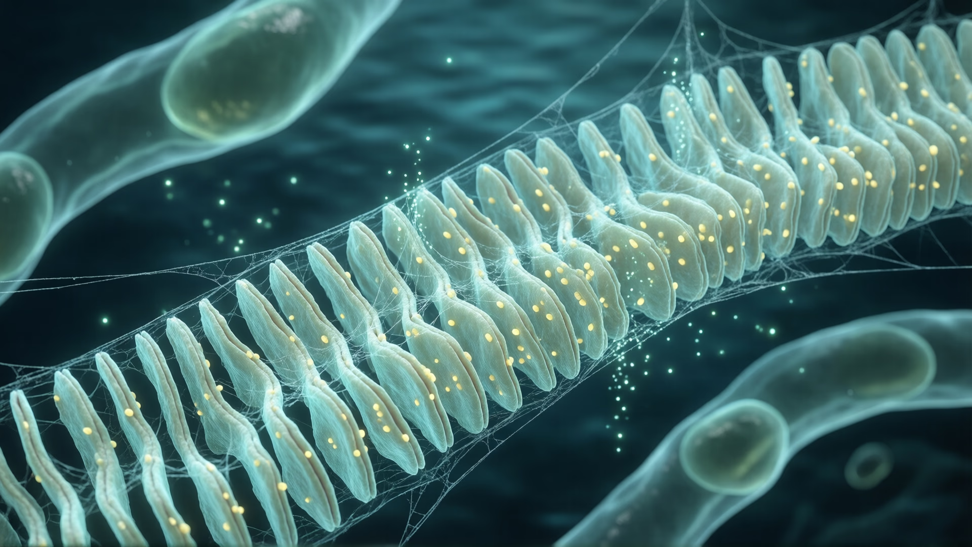
The endoplasmic reticulum is characterized by its continuous membranous structure, which connects with the nuclear envelope and plasma membrane. It exists in two primary forms: rough endoplasmic reticulum (RER) and smooth endoplasmic reticulum (SER). The RER is studded with ribosomes responsible for protein synthesis, while the SER lacks ribosomes and is involved in lipid metabolism and other synthetic processes.
Membrane Characteristics
The ER membrane is approximately 50 to 60 angstroms thick and exhibits a fluid-mosaic structure similar to the plasma membrane. It contains essential enzymes such as stearases, NADH-cytochrome C reductase, and glucose-6-phosphatase, which are crucial for synthetic and metabolic functions.
Morphological Forms
The ER can be visualized in three distinct morphological forms:
- Lamellar (Cisternae): Long, flattened sac-like structures found in cells like pancreatic cells and brain cells.
- Vesicular: Membrane-bound vacuoles ranging from 25 to 500 micrometers in diameter, predominantly seen in the SER.
- Tubular: Branched tubules that connect with cisternae and vesicles, commonly observed in most eukaryotic cells.
Types of Endoplasmic Reticulum
Smooth Endoplasmic Reticulum (SER)
- Function: Lipid metabolism (phospholipids, cholesterol, and steroid synthesis), glycogenolysis, and drug detoxification.
- Enzymes: Cytochrome P-450 enzymes for detoxification.
- Cell Types: Adipose cells, liver cells, heart conduction fibers.
Rough Endoplasmic Reticulum (RER)
- Function: Protein synthesis and maturation. It is the site of cotranslational protein synthesis, where ribosomes attached to its surface synthesize polypeptides that are inserted into the ER lumen for folding and modification.
- Characteristics: Contains transmembrane glycoproteins called ribophorins I and II.
- Cell Types: Pancreatic cells, plasma cells, liver cells.
Functions
The ER performs diverse functions critical to cellular activity:
- Lipid Metabolism: Smooth ER synthesizes phospholipids and steroids.
- Glycogenolysis: Breaks down glycogen into glucose units.
- Detoxification: Smooth ER detoxifies drugs through cytochrome P-450 enzymes.
- Mechanical Support: Provides structural support to the cell.
- Transportation: Acts as an intracellular transport system for molecules.
- Protein Maturation: Rough ER facilitates protein folding and modification, packaging proteins into vesicles that fuse with the Golgi apparatus.
- Signal Transmission: Plays a role in transmitting intra-cellular signals, such as muscle contractions.
- Endomembrane System: Integral part of the endomembrane system, including the nuclear envelope and Golgi apparatus.
Calcium Regulation
Calcium released from the ER is essential for various cellular processes such as muscle contraction, neurotransmitter release, and cell signaling. The IP3 receptor on the ER membrane facilitates this release, highlighting the ER's role in maintaining intracellular calcium homeostasis.
Signaling Pathways
The interaction between GPCRs and PLC leads to downstream effects mediated by IP3 and DAG. These molecules activate protein kinase C and promote calcium mobilization from the ER, illustrating the ER's involvement in intricate signaling mechanisms.
Role in Cellular Communication
The ER is integral to signaling pathways that regulate cellular responses. G-protein-coupled receptors (GPCRs) on the cell surface activate phospholipase C (PLC), which cleaves phosphatidylinositol-4,5-bisphosphate (PIP2) into inositol triphosphate (IP3) and diacylglycerol (DAG). IP3 binds to receptors on the ER, triggering the release of calcium ions into the cytoplasm.
Clinical Relevance
Disruptions in ER function can lead to pathological conditions. For instance, in Graves disease, autoantibodies binding to TSH receptors cause excessive thyroid hormone production, underscoring the importance of proper ER signaling in maintaining hormonal balance and cellular regulation.
[1]: Endoplasmic Reticulum (ER): Structure and Functions [2]: Physiology, Cellular Messengers - StatPearls - NCBI Bookshelf